Basal Ganglia's Crucial Role in Stroke Recovery


Intro
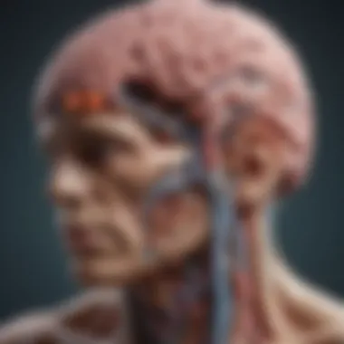
The basal ganglia are a group of nuclei in the brain that play a significant role in motor control, as well as various cognitive functions. Their involvement in stroke, a major global health concern, is profound. Understanding how the basal ganglia function, especially after a stroke, can help clarify the implications for recovery and rehabilitation.
When a stroke occurs, the blood supply to the brain is disrupted, leading to neuronal damage. This damage can affect many functions of the brain, including those governed by the basal ganglia. The complex interplay between the basal ganglia and stroke outcomes highlights the necessity to explore their role in the recovery process.
This article delves into multiple aspects of this relationship. Key points include the alterations in the basal ganglia post-stroke, the implications for patient rehabilitation, and avenues for future research. Furthermore, this discussion will emphasize neuroplasticity — the brain's ability to reorganize and adapt following injury — which is crucial for recovery.
The examination of risk factors and the mechanisms underlying stroke will provide insightful context for why certain patients might respond differently to rehabilitation strategies. Overall, understanding the intricate role of the basal ganglia in stroke is vital for advancing clinical practices and improving patient outcomes.
Preface to Basal Ganglia and Stroke
The relationship between the basal ganglia and stroke is crucial for understanding the broader implications of cerebrovascular accidents. This section serves as an entry point into the interconnectedness of these two topics, illustrating why they matter, especially in clinical practice and rehabilitation.
Stroke can have far-reaching impacts on brain function, and the basal ganglia play a prominent role in motor control and various cognitive processes. Understanding this relationship helps in recognizing the challenges that stroke survivors face, particularly in movement and coordination. Furthermore, the knowledge of how strokes affect the basal ganglia guides medical professionals as they develop effective treatment plans and rehabilitation strategies.
As such, a deep dive into the anatomy and function of the basal ganglia will provide essential insights into how stroke alters these neural circuits and the subsequent implications for recovery.
Defining Basal Ganglia
The basal ganglia consist of several interconnected structures that influence movement and coordination. These include the striatum, globus pallidus, substantia nigra, and subthalamic nucleus. Each of these components interacts intricately to modulate motor output and maintain voluntary movements.
The primary role of the basal ganglia centers around the initiation and regulation of movement. They help filter out unnecessary movements while facilitating the execution of intended actions. Dysfunction in this system can result in various movement disorders, which often complicate stroke recovery.
Understanding the configuration and operational dynamics of the basal ganglia is critical for grasping how strokes can disrupt these processes. This disruption can lead to a range of motor deficits, highlighting the importance of maintaining the integrity of these neural pathways.
Understanding Stroke
Stroke is a medical condition that occurs when the blood supply to a part of the brain is interrupted or severely reduced, preventing brain tissue from receiving necessary oxygen and nutrients. This can be classified mainly into two categories: ischemic and hemorrhagic strokes.
Ischemic strokes occur due to a blockage in a blood vessel, often caused by a clot. Conversely, hemorrhagic strokes result from a rupture in a blood vessel, leading to bleeding in or around the brain. Both types can lead to significant cerebral damage and impact the functioning of various brain structures, including the basal ganglia.
The clinical manifestations of a stroke vary widely, depending on the location and extent of the brain injury. Common symptoms include weakness or paralysis on one side of the body, difficulty speaking, and challenges with coordination. The prognosis and recovery potential depend heavily on the extent of the damage and timely intervention, underlining the need for rapid response in medical situations.
Anatomy of the Basal Ganglia
The anatomy of the basal ganglia is central to understanding its role in stroke and recovery. The basal ganglia consists of several nuclei that work together to facilitate movement control and coordination. Disruption in this system during a stroke can lead to significant impairments. A thorough exploration of its anatomical components is crucial for clinicians and researchers in addressing the functional repercussions of stroke.
Components of Basal Ganglia
Striatum
The striatum is one of the largest components of the basal ganglia. It is divided into two main parts: the caudate nucleus and the putamen. Its primary role in motor planning and execution positions it as a significant player in the overall topic. A key characteristic of the striatum is its receipt of direct input from the cerebral cortex, making it a beneficial area to examine in stroke research. Its unique feature is the integration of both motor and cognitive functions, which presents advantages in understanding how stroke affects various neural pathways and movements.
Globus Pallidus
The globus pallidus consists of two segments: the internal and external segments. It is essential in regulating voluntary movement by sending inhibitory signals to the thalamus and other motor centers. This area is particularly notable for its ability to modulate motor commands. The globus pallidus is also deemed beneficial for discussions around stroke, as its dysfunction can lead to movement disorders. Its unique feature lies in its complex interactions with other basal ganglia structures, leading to both advantages and disadvantages in understanding motor control intricacies post-stroke.
Substantia Nigra
The substantia nigra plays a vital role in the reward system and movement control. This area is key because it produces dopamine, which is crucial for smooth and coordinated muscle movements. The loss of dopaminergic neurons in this region is often linked to motor deficits observed in stroke patients. This unique characteristic makes it an important focus of recovery strategies. Its involvement carries certain disadvantages too, particularly in how dopaminergic loss can exacerbate recovery challenges.
Subthalamic Nucleus
The subthalamic nucleus is thought to play a role in regulating movements. It connects to various parts of the basal ganglia and contributes to the processing of motor signals. The key characteristic of the subthalamic nucleus is its excitatory influence on pathways that lead to movement facilitation. Studying this area can provide vital insights into stroke impact on motor functions. Its unique feature is its ability to enhance or dampen motor signals, presenting distinct advantages and disadvantages in the rehabilitation context.
Functional Significance
Understanding the anatomy of the basal ganglia allows for a fuller comprehension of its functional significance in stroke recovery. The coordination between these component structures establishes a comprehensive approach to analyzing motor outcomes. Regular examination of these aspects highlights the importance of tailored rehabilitation strategies for stroke patients.


Mechanisms of Stroke Impacting Basal Ganglia
The understanding of stroke mechanisms is crucial for grasping how the basal ganglia can be affected during a stroke event. The basal ganglia play significant roles in motor control and various cognitive functions. Their impairment can have profound effects on recovery and rehabilitation. A nuanced exploration of ischemic and hemorrhagic strokes illustrates how these conditions can lead to distinct types of neurological damage, thus informing treatment protocols.
Ischemic vs. Hemorrhagic Stroke
Ischemic strokes occur when an artery supplying blood to the brain is blocked. This can happen due to a blood clot or narrowing of the arteries. In contrast, hemorrhagic strokes are caused by the rupture of a blood vessel, which leads to bleeding in or around the brain.
Understanding these differences is vital:
- Ischemic Stroke: Often results in cell death in the areas deprived of blood, impacting the basal ganglia's function and leading to motor deficits.
- Hemorrhagic Stroke: The immediate damage from bleeding creates pressure in the brain, often causing a more severe and sudden impact on the basal ganglia than ischemic strokes.
Both types of strokes affect neurotransmitter systems and blood flow in the basal ganglia. The specific injury mechanism influences the potential for recovery, making it essential to adapt rehabilitation strategies accordingly.
Neurological Damage and Recovery
The extent of neurological damage in the basal ganglia post-stroke heavily depends on the type and severity of the stroke. Following an ischemic stroke, the immediate loss of blood supply leads to damaged neurons and the surrounding tissue. In hemorrhagic strokes, the damage may also be compounded by increased intracranial pressure and secondary injury.
Recovery after stroke varies significantly among individuals:
- Neuroplasticity: The brain's capacity to reorganize itself plays a crucial role in recovery. Neuroplasticity allows undamaged regions to take over some functions of the affected areas. However, reliance on neuroplasticity may not always compensate for significant basal ganglia damage.
- Rehabilitation: Effective rehabilitation depends on the type of stroke and the patient’s current abilities. Engaging in therapies that target motor skills can promote recovery by encouraging the brain to forge new connections.
"Rehabilitation strategies should be tailored according to the particular needs of the patient after a stroke, emphasizing the role of the basal ganglia in movement control."
The understanding of these neurological processes and their implications can lead to more effective clinical assessments and improved recovery outcomes for stroke patients.
This segment serves to highlight the intricate relationship between the mechanisms of stroke and the critical role of basal ganglia, paving the way for future research and clinical practice.
Role of Basal Ganglia in Motor Control Post-Stroke
The role of the basal ganglia in motor control post-stroke is crucial for understanding the recovery process. These structures in the brain are involved in refining movements and facilitating smooth motor function. When a stroke occurs, the communication pathways that involve the basal ganglia may be disrupted. This disruption leads to various motor function challenges, impacting the patient's ability to perform daily tasks. Recognizing the significance of the basal ganglia helps clinicians and caregivers to identify specific impairments and tailor rehabilitation strategies effectively.
Motor Function Impairments
Motor function impairments are common after a stroke. Patients may experience weakness or paralysis on one side of the body, known as hemiplegia. The basal ganglia contribute to the control of voluntary movements, and damage to this region can result in a lack of coordination.
Common impairments include:
- Weakness: Reduced strength in the arm or leg on the affected side.
- Rigidity: Stiffness in muscles, making movements difficult.
- Bradykinesia: Slowness of movement that affects speed and agility.
- Dystonia: Involuntary muscle contractions leading to abnormal postures.
Understanding these impairments is essential for developing appropriate rehabilitation strategies that focus on restoring motor function. The involvement of the basal ganglia underlines the need for targeted therapies aimed at improving motor control.
Movement Disorders Following Stroke
Stroke can lead to several movement disorders linked to the dysfunction of the basal ganglia. The interplay between the basal ganglia and other brain regions can be disrupted, resulting in a variety of movement issues.
Some of the common disorders include:
- Parkinsonism: Symptoms may mimic Parkinson's disease, with trembling, stiff limbs, and trouble initiating movement.
- Hemiballismus: A rare condition where one side of the body experiences sudden, wild movements. It occurs due to lesions in the subthalamic nucleus, part of the basal ganglia.
- Athetosis: Characterized by slow, writhing movements, often affecting the hands and feet.
These disorders complicate the recovery process. Understanding these conditions helps in diagnosing the exact issues experienced by stroke patients. Clinicians can utilize this knowledge to design individualized treatment programs that address specific challenges in movement control.
"Rehabilitation is not just about physical recovery; it is the journey of reclaiming one’s motor abilities and independence post-stroke."
Neuroplasticity and Basal Ganglia After Stroke
The concept of neuroplasticity is essential in the context of stroke recovery. Neuroplasticity refers to the brain's ability to reorganize itself by forming new neural connections. This occurs in response to learning, experience, or injury. After a stroke, the basal ganglia, which play a critical role in the coordination and regulation of movement, are often affected. Understanding how neuroplasticity works within the basal ganglia following a stroke can provide insights into therapeutic strategies and rehabilitation techniques.
Neuroplasticity offers several benefits for stroke patients. It enables the recruitment of undamaged brain areas to compensate for lost functions. Additionally, it supports the restoration of motor control and cognitive functions through intensive therapy and practice. The basal ganglia interact with other brain regions, facilitating this adaptation. By leveraging neuroplastic changes, rehabilitation can be more effective, leading to a better recovery outcome.


However, there are considerations to keep in mind. The extent of neuroplasticity varies among individuals, influenced by factors such as age, initial stroke severity, and pre-existing health conditions. Thus, tailored rehabilitation approaches are vital.
Concept of Neuroplasticity
Neuroplasticity is fundamentally about change. It is the brain’s ability to alter its structure and function in response to new information or injury. After a stroke, the damaged areas can lead to deficits in motor skills, speech, and other functions. The good news is that the brain can form new connections, allowing for recovery.
There are two main types of neuroplasticity: functional and structural. Functional plasticity is when the healthy areas of the brain take over the functions of the damaged ones. Structural plasticity involves physical changes at the synaptic level, such as the growth of new neurons or dendrites.
Rehabilitation Techniques Promoting Recovery
Different rehabilitation techniques are pivotal in promoting neuroplasticity and recovery after stroke. Understanding these methods helps in recognizing their unique contributions.
Physical Therapy
Physical therapy focuses on improving mobility and physical function. It often includes exercises designed to strengthen muscles and enhance coordination. The key characteristic of physical therapy lies in its ability to provide a structured environment for movement practice.
This discipline is widely regarded as a beneficial choice because it directly targets physical limitations caused by stroke. Unique features include task-specific training, where patients practice specific movements related to daily activities.
While physical therapy is generally advantageous, it can be demanding. Patients may experience frustration during recovery, as progress can be gradual.
Occupational Therapy
Occupational therapy aims to help stroke patients regain independence in daily activities. This type of therapy emphasizes the adaptation of tasks and environments to suit the patient’s abilities. One key characteristic is its focus on real-life context and meaningful activities. This makes it a popular choice among rehabilitative methods.
Occupational therapy often incorporates adaptive devices which assist patients in carrying out daily tasks. However, one of the disadvantages is that progress can vary widely between patients, making it hard to set clear recovery timelines.
Speech Therapy
Speech therapy addresses communication and swallowing difficulties resulting from a stroke. This area is crucial for patients as these functions are often impacted significantly. The main characteristic of speech therapy is its focus on restoring verbal and non-verbal communication skills.
Speech therapy is beneficial because it can increase patient participation in social settings, improving their quality of life. A unique feature of this therapy is the use of various techniques, such as exercises for articulatory precision, vocal training, and language skills enhancement. Challenges can arise, however, as not all patients respond equally, and some may experience longer recovery times.
Understanding and applying neuroplasticity principles in therapy can greatly enhance recovery outcomes for stroke survivors.
Assessment and Diagnosis of Post-Stroke Conditions
Assessing and diagnosing post-stroke conditions is a crucial aspect of managing stroke recovery. This phase determines the extent of neurological damage and impacts rehabilitation outcomes. Accurate assessments are foundational for tailoring treatment plans that effectively address the specific needs of the patient. Moreover, understanding the patient's condition helps inform caregivers about potential complications and promote better quality of life.
Diagnostic Imaging Techniques
CT Scans
CT scans play a vital role in the early evaluation of stroke patients. They provide rapid imaging that can help distinguish between ischemic and hemorrhagic strokes, which is essential for effective management. One of the key characteristics of CT scans is their speed. They can be performed quickly, which is critical in emergency settings. The unique feature of CT scans is their ability to visualize acute bleeding in the brain. This advantage allows for immediate interventions when there is a hemorrhagic stroke.
However, CT scans have some disadvantages. They may not detect smaller ischemic strokes, and repeated exposure to radiation can be concerning, particularly for vulnerable populations such as children and the elderly. In summary, while CT scans offer valuable insights in time-sensitive situations, they must be complemented by other imaging modalities in certain cases.
MRIs
MRIs are increasingly used in the assessment of post-stroke patients. Their high-resolution images allow for a detailed view of brain structures and abnormalities. A key characteristic of MRIs is their ability to provide more comprehensive information about cerebral tissue, including subtle differences between healthy and damaged areas. This makes them particularly useful in identifying smaller lesions that may go undetected by CT scans.
Clinical Assessments
Clinical assessments are essential tools for evaluating the functional status of stroke patients. These assessments help identify specific deficits that affect daily living and guide rehabilitation efforts. Their comprehensive nature ensures that the treatment plans are customized to address individual patient needs.
Neurological Exams
Neurological exams are a foundational aspect of clinical assessments in stroke management. They evaluate various cognitive and motor functions, offering a snapshot of the patient's current capabilities. The key characteristic of neurological exams is their ability to quickly identify critical changes in a patient’s condition. This immediacy makes them a beneficial choice, especially during hospital admissions.
A unique feature of neurological exams is that they can be performed at the bedside, minimizing the need for complex equipment. However, one disadvantage is that they rely significantly on clinician expertise. Interpretation can vary, influencing the consistency of results. Overall, neurological exams are indispensable for immediate assessments but must be conducted by trained professionals to ensure accuracy.


Functional Scales
Functional scales provide structured ways to assess the ability of post-stroke patients to perform daily activities. They measure various performance levels and can help track changes over time. A key characteristic of functional scales is their standardized scoring systems, which facilitate comparisons across different studies and populations. This standardization makes them a popular choice in stroke research and rehabilitation settings.
One unique aspect of functional scales is their focus on activities of daily living, which are essential for patient autonomy. The downside is that they may not capture all aspects of a patient's condition, especially in cases where cognitive deficits are prominent. Consequently, while functional scales provide valuable insights, they should be used in conjunction with other assessment tools to capture a more comprehensive view of a patient's recovery.
In stroke management, combining various diagnostic and clinical assessment techniques yields a holistic understanding of post-stroke conditions.
Pharmacological Approaches to Stroke Management
Pharmacological approaches play a crucial role in stroke management. These strategies aim to minimize damage caused by the stroke and enhance recovery. The careful selection and application of medications can significantly influence patient outcomes, and understanding these approaches is essential for healthcare providers and researchers alike.
Thrombolytic Therapy
Thrombolytic therapy is often one of the first lines of treatment for patients suffering from ischemic stroke, where a clot blocks blood flow to the brain. This therapy involves the administration of drugs that dissolve blood clots, restoring blood circulation. One common agent used is alteplase, also known as tissue plasminogen activator (tPA). The effectiveness of thrombolytic therapy is time-sensitive; it is most effective when initiated within a few hours of symptom onset.
The main benefits of this therapy include:
- Rapid relief of clot obstruction: Fast restoration of blood flow is critical to minimizing brain damage.
- Improved clinical outcomes: Patients who receive timely thrombolytic treatment often show better recovery results compared to those who do not receive it.
- Potential to reduce disability: By promoting quicker recovery, thrombolytic therapy may decrease long-term disabilities often associated with stroke.
However, there are also important considerations and risks associated with thrombolytic therapy:
- Risk of hemorrhage: The administration of thrombolytics increases the risk of bleeding, which can lead to further complications.
- Need for careful patient selection: Not all patients are candidates for thrombolytic therapy. Factors such as the type of stroke, overall health status, and timing of treatment must be evaluated.
Neuroprotective Agents
Neuroprotective agents focus on protecting neuronal cells from damage during and after a stroke. These medications are still under investigation but hold considerable promise for enhancing recovery. The aim is to provide a defensive mechanism against the resultant cascade of biochemical events that lead to cell death post-stroke.
Some potential neuroprotective agents include:
- N-Methyl-D-Aspartate (NMDA) receptor antagonists: These can reduce excitotoxicity, which occurs when over-stimulation of neurons leads to cell injury or death, a common issue following stroke.
- Calcium channel blockers: These help to prevent excessive calcium influx into cells, which can result in cell injury.
- Free radical scavengers: These agents combat oxidative stress, another damaging process following a stroke.
Benefits of using neuroprotective agents are promising:
- Reduction of cell death: Protecting neurons may help preserve brain tissue and function.
- Support for rehabilitation: When coupled with rehabilitation efforts, neuroprotective drugs might enhance the recovery process.
Nonetheless, it is essential to be aware that the use of neuroprotective agents is primarily experimental at this point. Ongoing clinical trials continue to explore their efficacy and safety.
"The pharmaceutical landscape for stroke management is evolving, and both thrombolytic therapy and neuroprotective agents are critical aspects to monitor."
In summary, pharmacological approaches are vital in stroke management. Thrombolytic therapy provides a time-sensitive intervention to restore blood flow, while neuroprotective agents offer a protective mechanism for the brain. Understanding and integrating these treatments can significantly impact patient recovery and quality of life.
Emerging Research and Future Directions
Emerging research related to the basal ganglia and stroke is vital in understanding how to improve recovery outcomes. The exploration of innovative approaches to therapeutic strategies can uncover ways to enhance neuroplasticity, which is essential for stroke rehabilitation. The advancements in the understanding of the basal ganglia's role in motor and cognitive functions post-stroke hold significant implications for clinical practices and patient care. New studies are increasingly focusing on personalized treatment plans that address individual patient needs based on their specific deficits and recovery potential.
Research in this field is characterized by a multidisciplinary approach, integrating neurology, rehabilitation science, and molecular biology. This interaction among different scientific areas promotes the development of effective interventions. One of the major benefits of focusing on emerging research is the potential for novel therapies that can make a meaningful difference in recovery outcomes. For instance, understanding how to manipulate neuroplasticity could lead to enhancements in existing rehabilitation strategies, making them more effective and tailored to each patient.
From a practical standpoint, it is essential for researchers to consider not just the biological mechanisms but also the social implications of their findings. Studies demonstrating innovative therapies must connect with health care providers and patient advocacy groups to ensure the research translates into practice.
Innovative Therapeutic Strategies
A focus on innovative therapeutic strategies is crucial. There is promising research on the use of robotics and technology to facilitate rehabilitation. Wearable devices and robotic exoskeletons can provide patients with feedback and assistance during their recovery processes. These technologies aim to engage patients actively, supporting them in completing movements and regaining motor functions. In addition, virtual reality environments are emerging as tools for rehabilitation, enabling repeatable practice of motor tasks in a controlled setting.
Such technologies combine traditional methods with advanced techniques. They can potentially lead to faster recovery times and improved outcomes. Furthermore, employing approaches like constraint-induced movement therapy has gained traction as it encourages the use of affected limbs, thus promoting neuroplastic changes within the basal ganglia.
Genetic and Molecular Studies
The exploration of genetic and molecular aspects related to stroke recovery is gaining momentum. Studies in this domain can elucidate how genetic predispositions impact individual recovery trajectories. Identifying specific genetic markers associated with stroke recovery can lead to more tailored therapeutic interventions.
For example, understanding the expression of certain genes involved in neuroinflammation may provide insights into potential interventions for mitigating neuronal death in the basal ganglia. Overall, ongoing genetic studies are vital in uncovering the biological underpinnings of recovery.
In summary, the future direction of research focusing on the basal ganglia and stroke recovery is quite promising. With advancements in both therapeutic strategies and genetic studies, there lies significant potential to improve clinical outcomes for stroke patients.
"Continued research into the intersections of genetics, rehabilitation technology, and neuroplasticity will pave the way for breakthroughs in stroke recovery therapies."
Engaging in this evolving field not only enhances our scientific understanding but also holds the potential to transform how patients recover from strokes.


